
By ECRI

Mobile radiographic units are used to perform radiographic studies at the point of care. When X-rays are needed, these machines are wheeled to the patient, rather than transporting the patient to the radiology department. Portable radiography is seen as a way to help reduce the spread of COVID-19 by bringing care to the patient; thus its use has increased during the pandemic. ECRI addressed this trend in its lab webcast series and device evaluations.
From his days practicing emergency medicine, ECRI’s Andrew Furman, MD, knows the value of being able to bring X-ray technology to the patient: “Physicians like the convenience of being able to take a quick snapshot of what’s happening with the patient,” Furman explains. But that’s not the only factor to consider: “A patient may be better served by a two-view study conducted in the radiology department. So, you weigh the options.”
One interesting aspect of practicing health care in the presence of COVID-19, Furman observes, is that “those risk-benefit calculations have changed.” Now, care providers are even more likely to decide that the risks associated with transporting the patient to the radiology department – specifically, the risks of spreading the SARS-CoV-2 virus – outweigh the benefits. That assessment makes portable radiography an even more valuable tool, a development explored during a November 2020 ECRI webcast hosted by Furman in his role as ECRI’s executive director of clinical excellence and technology assessment.
The webcast panel discussed how to safely adapt radiographic services during the COVID-19 pandemic, strategies for conserving personal protective equipment (PPE) during mobile imaging studies, and operational and image quality considerations with portable radiography. Also discussed were key findings from ECRI’s testing of mobile radiographic units. (Members of certain ECRI programs can access a recording of the webcast, “Key Considerations for Portable Radiography in the Era of COVID-19,” as well as ECRI’s evaluation findings.)
Mobile Radiographic Units: The Basics
Mobile radiographic units are used to image patients for whom transport is contraindicated. Traditionally, these machines have been used in areas such as intensive or critical care units, operating rooms, pediatric areas and emergency rooms. In the past year, their use has expanded to include areas where known or suspected COVID-19 patients are being treated.
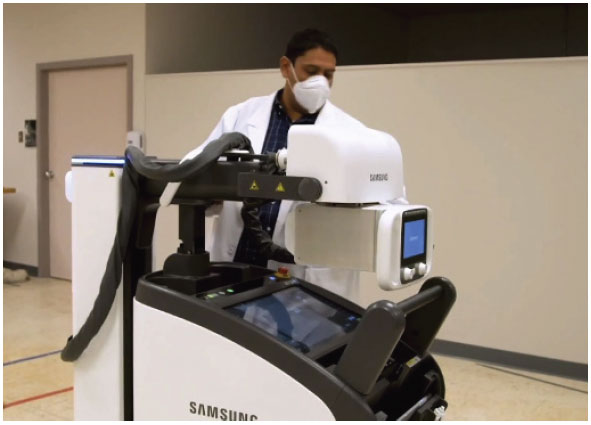
These devices are self-contained, battery-operated motorized carts with a telescopic column. An X-ray generator provides power to the device’s X-ray tube, which generates the X-ray beam. Movement of the machine is facilitated by highly maneuverable motor drives; the drives, which are controlled by a handlebar equipped with pressure sensors, allow the unit to perform a zero-radius turn, and they support fine-positioning movements to help set up the exam. A computer provides a graphical user interface and tools for selecting the patient, setting up the exam parameters, acquiring the image, reviewing and annotating the image, and sending the image to an archiving system. Additionally, the systems ECRI has tested have wireless flat-panel detectors for acquiring the images.
When an exam is ordered, the technologist drives the unit to the patient, positions it at the bedside, places the detector behind the area of interest on the patient, selects the X-ray parameters and acquires the images. The images are displayed within a few seconds on the computer display. If the images are acceptable, they are sent to the PACS for reading by a radiologist.
Key Selection Factors in the Era of COVID-19
ECRI has tested a dozen mobile radiographic units in recent years. All the systems provide good image quality and deliver the necessary clinical functionality. Often, selection decisions will come down to differences in a few performance factors and workflow features that affect ease of use. A few of these factors warrant greater emphasis in a COVID-19 world:
- Battery capacity: High battery capacity allows the user to perform a large number of exams without any delays caused by the need to charge the battery between exams. During testing, ECRI measures the number of exposures that were possible in a simulated workflow. “Battery life is always an issue, but it’s an even bigger consideration now,” remarks Francisco Rodriguez-Campos MSc, Ph.D., MRSO (MRSC™), a senior project officer in ECRI’s device evaluation group. “You may be taking the machine outside the normal area of operation, or using it longer than previously, so you’ll want to know that the battery will last for the shift.”
- Size and weight of the cart and the detectors: Smaller and lighter systems are easier to control, maneuver and position. Similarly, detectors that are lightweight and appropriately sized can more easily and quickly be positioned to acquire images for various applications. “These days, you want to minimize interactions near the patient to protect staff from infectious disease exposures,” explains Rodriguez-Campos.
- Positioning tools and features: Fine-positioning tools improve ease of use and help shorten exam setup time. When imaging known or suspected COVID-19 patients, ease of positioning the tube head for an exam, for example, takes on added significance.
- Cleaning and disinfection: With mobile radiographic units being moved from one potentially infectious patient to another, frequent cleaning and disinfection of the unit is essential. Consider the unit’s IPX rating, which describes its resistance to fluid ingress, and verify its compatibility with various cleaning supplies. The use of incompatible cleaning and disinfection solutions can cause expensive damage to the unit.
Other performance and workflow factors, such as the following, will always warrant consideration:
- X-ray generator power rating: Sufficient power is essential to obtain an adequate-quality image. Higher output power is an advantage: It allows shorter exposures, thereby reducing the effect of patient movements that degrade image quality.
- X-ray tube heat capacity and cooling rate: Considerable heat is generated during X-ray production; thus the maximum heat that the X-ray tube can withstand and the maximum cooling rate should be considered together. The X-ray system will automatically limit the exposure options if the heat generated by an exposure would be too high. To avoid delays, it is important to have both a high heat capacity and a high cooling rate.
- Ability to make exposures using AC power: All units are designed to make exposures using the battery. Some can make an exposure when plugged into to a standard outlet, which is an advantage when the battery becomes depleted.
This article is adapted from ECRI’s Evaluation Background: Mobile Radiographic Units with Wireless Digital Detectors. The complete article – including model-specific test results and product ratings, along with additional guidance for purchasing and using mobile radiographic units – is available to members of ECRI’s capital purchasing and device evaluation programs. To learn more about ECRI membership, visit www.ecri.org/solutions/technology-decision-support, or contact ECRI by telephone at 610-825-6000, ext. 5891, or by email at clientservices@ecri.org.
