

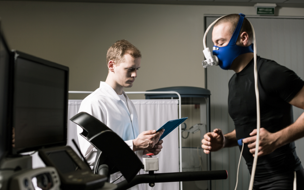
 Clinicians evaluate an individual’s response to physical stress through two primary types of stress exercise systems: cardiac systems, which primarily analyze electrocardiograms (ECGs), and pulmonary systems, which analyze respiratory gases/volumes and other parameters of pulmonary function.
Some stress exercise systems can provide both types of data by interfacing with peripheral devices; for example, some cardiac stress testing systems interface with respiratory gas analyzers, providing information about carbon dioxide (CO2) and oxygen (O2) utilization, as well as ECG data.
The ECG in exercise stress testing differs from a resting ECG in the length of the monitoring phase and in the parameters measured. Typically, a resting ECG measures 15 to 30 seconds of the heart’s electrical activity; stress exercise test protocols monitor the patient’s ECG for longer periods under different degrees of exercise. The ECG data is analyzed; reports can be generated from the system’s printer.
Before beginning the cardiac stress test, the clinician typically performs a three-phase pretest ECG protocol: ECG readings are taken with the subject supine, standing at rest, and hyperventilating. The purpose of the pretests is to provide baseline data to compare with the ECG during the stress exercise test.
The subject then begins the stress test according to a predetermined protocol that gradually increases the speed and/or grade of the treadmill exercise or the work performed with the ergometer. Most systems are preprogrammed with standard exercise protocols, such as Bruce, Balke, and Naughton, and can also be easily programmed by the user for a physician’s personal protocol. Each user can individualize the system by programming the frequency of blood pressure prompts, ST-segment measurement criteria, and leads displayed. Some systems allow the user to design the entire screen display to the clinician’s preference. Protocols can be modified with manual override for individual patients. Final report formats can include printing raw data and summarizing the activity of the ST segment and arrhythmias.
Some systems use a Microvolt T-Wave Alternans test to identify patients with life-threatening heart rhythms. This test requires an elevated heart rate; a treadmill is typically used. The clinician applies electrodes to the patient, which are then connected to the Microvolt T-Wave Alternans equipment. This test measures the T-wave at a microvolt level allowing clinicians to observe variations of the T-wave that can be indicators for risk of sudden cardiac death.
Pulmonary stress testing systems use O2 and CO2 analyzers to measure respiratory gases during exercise. For most systems, O2 concentration is measured by either an electrochemical or a paramagnetic method. Electrochemical methods include the use of a solid state electrolyte O2 sensor that uses zirconia stabilized with yttrium and the use of aqueous electrolyte electrochemical cells. Paramagnetic methods rely on the high magnetic susceptibility of O2 molecules. When drawn into the paramagnetic analyzer’s cell, the O2 causes a change in an applied magnetic field that is detected by the sensor. These various sensors can be used to measure the partial pressure of O2 in the sample.
CO2 analyzers for stress exercise testing typically use the principle of infrared (IR) absorption spectroscopy to measure the concentration of CO2 in expired gas. CO2 and water vapor selectively absorb specific wavelengths of IR light, making it possible to differentiate these gases from other commonly respired gases. Because the amount of IR light absorbed is proportional to the absorbing molecule’s concentration, the sample’s CO2 concentration can be determined by comparing its absorbance to that of a standard of known concentration.
Pulmonary stress exercise systems use either breath-by-breath analysis or mixing-chamber analysis. In breath-by-breath analysis, gases are measured and calculated from each breath as the subject exercises. In mixing-chamber analysis, several breaths are collected in a mixing chamber, and an average output of these breaths is used to calculate gas exchange. Breath-by-breath systems typically use a mask or mouthpiece attached to tubing that is connected to the gas analyzers; mixing-chamber systems use a mouthpiece, mask, or hood to obtain samples.
There are three basic types of pulmonary stress exercise protocols: steady-state testing, in which the subject stays at one workload for five minutes, while O2 and CO2 exchange is evaluated during the last minute; incremental testing, in which the clinician increases the workload each minute by 15 to 25 watts until a predetermined work goal is reached; and ramp testing, in which work is increased gradually and continuously until the patient can no longer perform.
Some cardiac and pulmonary stress exercise testing systems may link to ECG or cardiology data management systems, which electronically store results for patient record keeping and for statistical analysis.
Reported Problems
Because clinicians often use standardized exercise testing for patients with compromised cardiovascular and pulmonary capabilities, the stress induced during testing can cause life-threatening disturbances that require immediate attention. Appropriate personnel and equipment for emergency resuscitation should always be available, and patients should be monitored closely to ensure that they are not overtaxed.
Other Considerations
Before purchasing a stress exercise system, the prospective buyer should analyze the clinical setting’s present and future requirements regarding the projected volume of stress testing, patient diagnoses, and method of record keeping. The specific features of these systems vary; therefore, each model’s specifications should be carefully evaluated. Users should ensure that the cost of disposables, electrodes, recording paper, filters, calibration gases, spirometers, breathing circuits) is identified before purchasing the system. Discounts may be available when a disposables contract is included in the original purchase.
Some suppliers offer upgrade packages that allow the user to adapt or improve an existing system at a lower cost than that of a completely new system.
This article is adapted from ECRI Institute’s Healthcare Product Comparison System (HPCS), a searchable database of technology overviews and product specifications for capital medical equipment. The source article is available online to members of ECRI Institute’s HPCS; learn more at www.ecri.org/components/HPCS
